


Home / Training / Manuals / Atlas of breast cancer early detection / Cases
Atlas of breast cancer early detection
Go back to the list of case studies
.png) Click on the pictures to magnify and display the legends
Click on the pictures to magnify and display the legends
| Case number: | 184 |
| Age: | 70 |
| Clinical presentation: | Postmenopausal woman with average risk of developing breast cancer presented with a left breast lump. Examination revealed a hard lump in the outer quadrant of the left breast. |
Mammography:
 |  |
 |  |
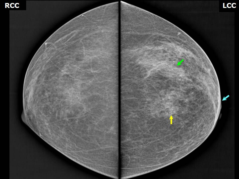
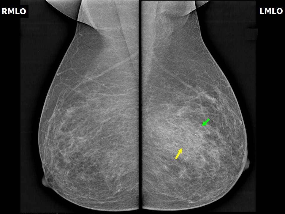
| Breast composition: | ACR category a (the breasts are almost entirely fatty) | Mammography features: |
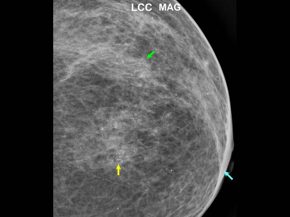
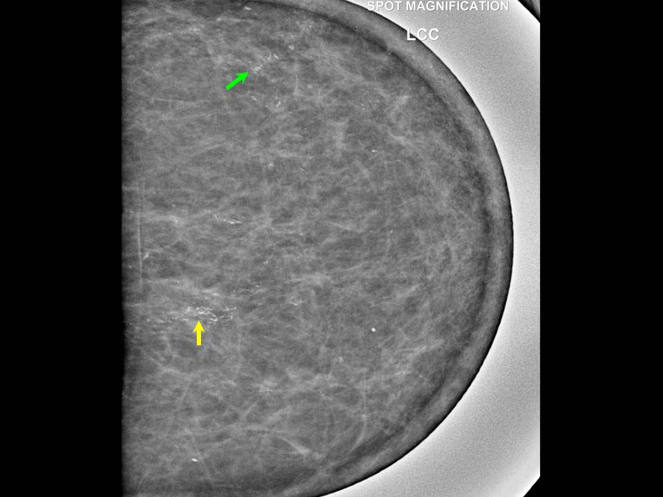
| ‣ Location of the lesion: | Left breast, upper outer quadrant at 122 oclock, anterior and middle thirds |
| ‣ Mass: | |
| • Number: | None |
| • Size: | None |
| • Shape: | None |
| • Margins: | None |
| • Density: | None |
| ‣ Calcifications: | |
| • Typically benign: | None |
| • Suspicious: | Fine pleomorphic microcalcifications in segmental distribution and in clusters and in linear branching pattern |
| • Distribution: | None |
| ‣ Architectural distortion: | None |
| ‣ Asymmetry: | Present |
| ‣ Intramammary node: | None |
| ‣ Skin lesion: | None |
| ‣ Solitary dilated duct: | None |
| ‣ Associated features: | Skin thickening |
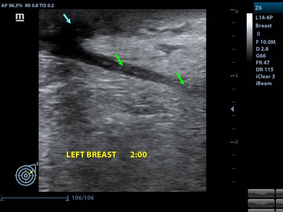
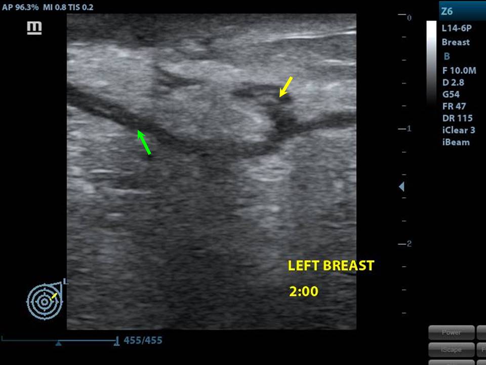
Ultrasound:
 |  |
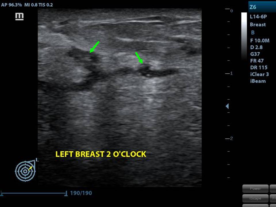 | 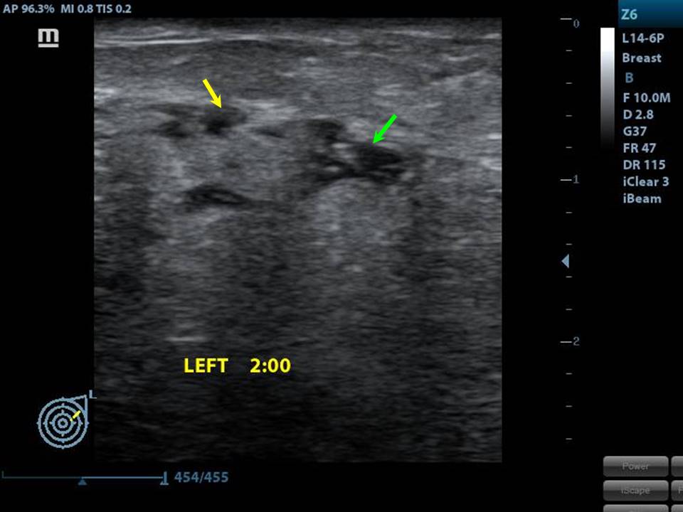 |
| Ultrasound features: Left breast, upper outer quadrant at 122 oclock | |
| ‣ Mass | |
| • Location: | Left breast, upper outer quadrant at 122 oclock |
| • Number: | Multiple small |
| • Size: | < 1 cm |
| • Shape: | Irregular |
| • Orientation: | Not parallel |
| • Margins: | Indistinct and angular |
| • Echo pattern: | Heteroechoic |
| • Posterior features: | No posterior features |
| ‣ Calcifications: | Intraductal microcalcifications |
| ‣ Associated features: | Axillary lymphadenopathy |
| ‣ Special cases: | Solitary dilated duct |
BI-RADS:
BI-RADS Category: 5 (highly suggestive of malignancy)Further assessment:
Further assessment advised: Referral for cytologyCytology:
 |
| Cytology features: | |
| ‣ Type of sample: | FNAC |
| ‣ Site of biopsy: | |
| • Laterality: | Left |
| • Quadrant: | Upper outer quadrant |
| • Localization technique: | Palpation |
| • Nature of aspirate: | Whitish, blood tinged |
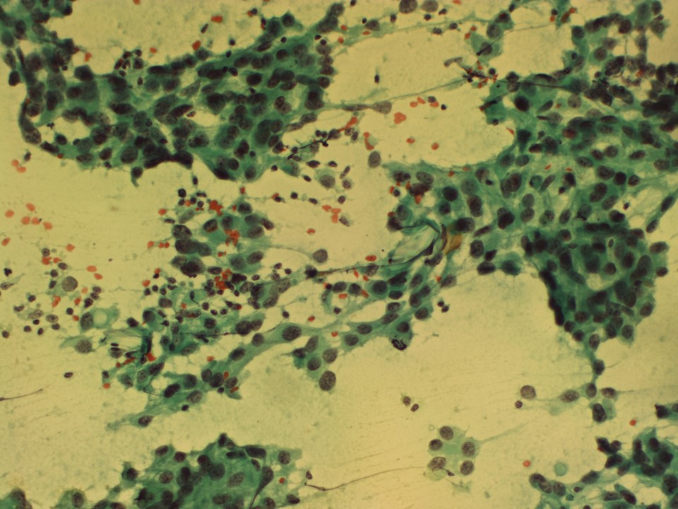
| ‣ Cytological description: | Smears are very cellular and show dyscohesive clusters of pleomorphic malignant cells |
| ‣ Reporting category: | Malignant |
| ‣ Diagnosis: | Carcinoma breast |
| ‣ Comments: | None |
Histopathology:
MRM
 |  |
| Histopathology features: | |
| ‣ Specimen type: | MRM |
| ‣ Laterality: | Left |
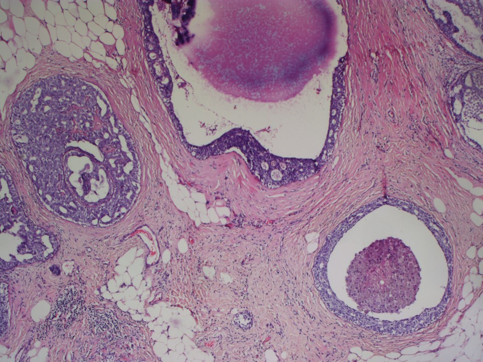
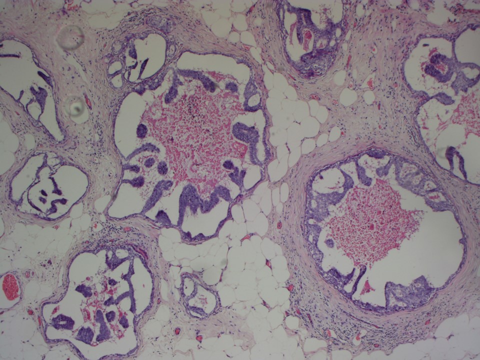
| ‣ Macroscopy: | Left MRM specimen (24.0 × 16.0 × 7.5 cm) with skin flap (11.0 × 6.5 cm). On serial sectioning, a firm greyish white area (6.0 × 3.0 × 3.0 cm) is seen in the upper outer quadrant and extending to the lower outer quadrant. A second firm area (1.5 × 1.0 × 1.0 cm) is seen 3 cm medial to the first firm area in the upper inner quadrant. The intervening breast parenchyma shows interspersed whitish calcified areas |
| ‣ Histological type: | Microinvasive breast carcinoma |
| ‣ Histological grade: | Grade 2 (3 + 2 + 2 = 7) |
| ‣ Mitosis: | 12 |
| ‣ Maximum invasive tumour size: | Six foci of microinvasive breast carcinoma; all < 0.1 cm in greatest dimension |
| ‣ Lymph node status: | 0/10 |
| ‣ Peritumoural lymphovascular invasion: | Absent |
| ‣ DCIS/EIC: | DCIS of cribriform, comedocarcinoma, and solid type, high grade with necrosis in both the firm areas seen grossly. EIC present. Multiple foci of DCIS extending over 6 cm in greatest dimension |
| ‣ Margins: | Free of tumour |
| ‣ Pathological stage: | pT1miNo |
| ‣ Biomarkers: | ER positive, PR positive, and HER2 negative |
| ‣ Comments: | The adjacent breast also shows lobular mastitis with a chronic inflammatory infiltrate |
Case summary:
| Postmenopausal woman presented with a left breast lump. Diagnosed as solitary dilated duct with fine pleomorphic microcalcifications in segmental distribution and in clusters and in linear branching pattern, BI-RADS 5 on imaging, as breast carcinoma on cytology, and as microinvasive breast carcinoma pT1miN0 with EIC on histopathology. |
Learning points:
|
EIC is defined by the following criteria:
|